
Glare Spread Function (GSF) - Characterization of optical glare:
Human Glare Spread Function
Human Vision’s GSF or retinal Point Spread Function(PSF)
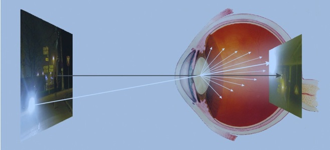
From: [1] Franssen L & Coppens JE (2007) “Straylight at the retina: Scattered papers”, Figure 1, thesis Universiteit van Amsterdam, Downloaded from UvA-DARE, the institutional repository of the University of Amsterdam (UvA)
http://hdl.handle.net/11245/2.46844
The above illustration of intraocular straylight is from L. Franssen and J. E. Coppens. This book provides an excellent introduction to retinal straylight (Chapter 2). It covers measurements of straylight in normal vision with variations due to age, and iris pigmentation. Further, it discusses straylight from a variety of classes of cataracts, straylight in the cornea. A comprehensive review can be found in “History of ocular straylight measurement: A review”[2], and “Ocular Media Clarity and Straylight”[3].
-
[2] van den Berg, T., Franssen, L., Bastiaan Kruijt, B., Coppens, J. E. (2013), “History of ocular straylight measurement: A review”, and “Ocular Media Clarity and Straylight”. Zeitschrift für Medizinische Physik, 23, 6-20.
http://dx.doi.org/10.1016/j.zemedi.2012.10.009
-
-
[3] van den Berg, T., Franssen, L., Coppens, J. E. (2010), “Ocular Media Clarity and Straylight “, in Encyclopedia of the Eye (2010), Academic Press, 3, 173-183. ISBN-9780123741981
The Effects of Glare
Retinal straylight is a visual handicap. Patient issues include hazy vision, contrast and color loss, difficulty with against-the-light face recognition, and halos around bright lights. Straylight will also adversely affect visual function tests, such as contrast sensitivity[4], visual field[5], and pattern electroretinogram [6].
CIE Standard Glare Observer
Cobb [7] introduced the concept of equivalent veiling luminance (Leq) as an apt way to define retinal straylight. Disability glare/retinal straylight, as defined by the CIE (Commission Internationale de l’Eclairage), is now quantified by means of this concept of equivalent luminance, i.e. the (external) luminance that has the same visual effect as the glare source at some angular distance.[8,9]
Leq is the outer part of the retinal point spread function PSF. The PSF is normalized to unity by writing
PSF = Leq/Ebl (sr -1)
with Ebl the illuminance on the eye from the (glare) point source.
Tom van den Berg, and J. K. IJspeert described a compensation technique to measure Leq/Ebl [10]. Vos, chair of CIE TC1-18 (Disability Glare) and van den Berg developed a Standard Glare Observer [11,12], accepted as CIE standard. [13]
Retinal Point Spread Function
Vos and van den Berg provided a detailed description of the shape of the retinal PSF in their 1999 “Report on disability glare”.[14] It include an equation with parameters for age and eye color. Using this retinal PSF one can calculate the retinal image of any well measured scene.
-----
-
[4] van den Berg, T. J. T. P. Importance of pathological intraocular light scatter for visual disability. Doc.Ophthalmol. 61(3-4), 327-333. 1-15-1986.
-
[5] van den Berg, T. J. T. P. Relation between media disturbances and the visual field. Doc Ophthalmol Proc Series 49, 33-38, 1987.
-
[6] van den Berg, T. J. T. P. and Boltjes, B. The point-spread function of the eye from 0 degrees to 100 degrees and the
pattern electroretinogram. Doc.Ophthalmol. 67(4), 347-354, 1987. -
[7] Cobb, P. W. The influence of illumination of the eye on visual acuity. Am J Physiol 29, 76-99, 1911.
-
[8] Vos, J. J. Disability glare - a state of the art report. Commission International de l'Eclairage Journal 3/2, 39-53. 1984.
-
[9] van den Berg, T. J. T. P. and IJspeert, J. K. Intraocular straylight, studied using the direct compensation technique. 22nd session(division 1), 83-84. 1991. CIE Proceedings.
-
[10] van den Berg, T. J. T. P. and IJspeert, J. K. Clinical assessment of intraocular straylight. Applied Optics 31, 3694- 3696. 1992.
-
[11] Vos, J. J. and van den Berg, T. J. T. P. Report on disability glare. CIE collection 135(1), 1-9. 1999.
-
[12] van den Berg, T. J. T. P. On the relation between glare and straylight. Doc.Ophthalmol. 78(3-4), 177-181. 1991.
-
[13] Vos, J. J., Cole, B. L., Bodmann, H-W., Colombo, E., Takeuchi, T., and van den Berg, T. J. T. P. CIE Equations for Disability glare. 2002. Commission Internationale d'Eclairage. CIE Collection on Glare.
-
[14] Vos, J. J. and van den Berg, T. J. T. P. Report on disability glare. CIE collection 135(1), 1-9. 1999.
Scene vs. Retinal Luminances
Numerous experiments have studied the relationship between luminance and sensation. White, gray, and black sensations, called achromatic lightnesses, or gray scales, have been studied by Munsell and many others.[12] In these studies lightnesses, the sensations generated by the visual system, are compared with relative luminances, measures of the amount of light coming from objects to the eye. We describe such measurements as luminance at the eyepoint.
A more relevant physical quantity for models of the visual psychophysics is the amount of light arriving at the receptors in the retina. By accounting for the physical effects of the scattering of light passing through the ocular media, one can calculated the relative stimulus at the retina. We convolve the luminance at the eyepoint with the retinal Point Spread Function (PSF) to calculate retinal luminance.
Calculating the Retinal Image
Stiehl et al.(1983) described an algorithm that calculated the contrast of selected pixels in an image on the human retina after intraocular scatter.[13] They calculated the luminance at selected pixels on the retina based on that display pixel’s measured luminance and the calculated scattered light from all other scene pixels. The calculation used Vos et al.’s (1976) measurements.[14] Stiehl measured the actual display luminance and calculated the retinal luminance.
Using Stiehl et al.’ s algorithm as a guide, Rizzi et al. [15,16,17] updated the process to use today’s more powerful imaging systems. They calculated the luminance on the retina based on the CIE standard for intraocular glare (Vos & van den Berg, 1999;[11] CIE, 2002;[10]. The goal was to compute the intensity of light falling onto the retina to separate the effects of glare from post-visual pigment neural contrast.
-----
-
[12] Wyszecki G. & Stiles WS (1982) “Colour Science: Concepts and Methods Quantitative Data and Formulae” , 2nd ed, John Wiley & Sons, Inc, New York, 486 – 513.
-
[13] Stiehl W. , McCann J. & Savoy R. (1983) Influence of Intraocular Scattered Light on Lightness-scaling Experiments, J Opt Soc Am, 73 , 1143 – 48.
-
[14] Vos J. J. Vos, J. Walraven, and A. van Meeteren, "Light profiles of the foveal image of a point source," Vision Res. 16, 215-219, (1976).
-
[15] Rizzi A , Pezzetti M & McCann J ( 2007 ) Glare - limited Appearances in HDR Images , IS & T/SID Color Imaging Conference , 15 , 293 – 8.
-
[16] Rizzi A. & McCann J. (2009) Glare- limited appearances in HDR images, J. Soc. Info. Display , 17 (1), 3 – 12 .
-
[17] McCann J. and A. Rizzi, (2012) “The Art and Science of HDR Imaging”, Wiley, Chichester, Ch 14-19.
-
ISBN: 978-0-470-66622-7